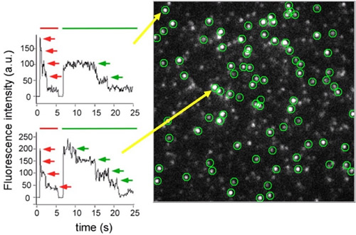
KCNQ1 and KCNE1 (both are known as causative genes of long QT syndrome) form an ion channel complex and regulate heart rhythm. To determine the stoichiometry of the complex, we directly counted the number of KCNQ1 and KCNE1 subunits by the newly-developed subunit counting method (Ulbrich, 2007). By counting spontaneous bleaching events of single GFP molecules tagged on these subunits using total internal reflection fluorescence (TIRF) microscope, we could determine how many GFP molecules (i.e. KCNQ1 or KCNE1 subunits) were included in one ion channel complex. Interestingly, the number of KCNE1 subunits in a single fluorescent spot was not fixed but flexible, and up to four KCNE1 subunits could bind to four KCNQ1 subunits (one ion channel). The average number of KCNE1 per one ion channel complex became higher when the relative expression density of KCNE1 against KCNQ1 was higher. As higher expression density of KCNE1 subunits made KCNQ1 channels harder to be open, the electrical activity of cardiac myocyte may be regulated by the relative expression densities of these subunits.
Nakajo K, Ulbrich MH, Kubo Y, Isacoff EY. (2010) Stoichiometry of the KCNQ1-KCNE1 ion channel complex. PNAS, 107: 18862-7
(Right) Representative image of GFP-tagged KCNE1 under the TIRF microscope is shown. Each green circle represents one ion channel complex. (Left) The time courses of the fluorescence intensity were shown. In the upper panel, two-time bleaching events of GFP were observed (green arrows), indicating that at least two GFPs (KCNE1 subunits) existed in the spot. Example of four GFPs (KCNE1 subunits) was shown in the lower panel.
*Div of Biophys and Neurobiol, NIPS


